TL;DR:
- Fish scales is a rare genetic skin condition (genodermatoses) characterized by excessive keratinization and layered, thick, scaly lesions.
- Symptoms include dryness, itching, irritation, limited movement, facial distortion, and risk of infection.
- Causes: Mutations in genes like KRT1, KRT10, KRT2, and FLG, disrupting keratin function and the skin's keratinization process.
- Types: Includes ichthyosis vulgaris, hedgehog fish scales, X-linked recessive variants, harlequin ichthyosis, ichthyotic erythroderma, bullous ichthyosis, and more.
- Diagnosis: Involves medical history, physical exams, skin biopsies, and genetic testing.
- Treatment: No cure exists. Symptomatic care includes moisturizers (with urea/salicylic acid), steroids, and possibly surgery. Ongoing specialist care is essential.
Fish scales is a rare, genetic skin disease that affects the appearance and function of the skin. It leads to the formation of thick, scaly lesions on the surface of the body. People with the condition struggle with dryness, itching and a tendency to irritation and infection. The condition has a significant impact on daily life and requires constant attention to skin care and lifestyle adjustments. In this article, we take a closer look at what fish scale is, the challenges it poses to sufferers, and what options are available.
What is fish scale?
Fish scales is a group of genetically determined diseases that are distinguished by a diverse clinical course. It belongs to a group of genodermatoses, the common feature of which is impaired keratinization and exfoliation of the skin. In general, the disorder leads to excessive keratinization of the epidermis and the formation of characteristically arranged scales that overlap in tile-like layers.
Clinical manifestations of fish scale.
Depending on the type of fish scale, skin lesions may appear all over the body or only in certain areas, including on the arms or legs. Excessive keratinization and desquamation leads to distorted facial features, curling of the lips and eyelids, and impaired facial expressions. On the other hand, ear cartilage dysfunction and excess keratinized skin can cause morphological changes in the auricles and narrowing of the external ear canals. Tight skin often leads to limited mobility in the upper and lower extremities and fingers. Tight skin bands, in turn, can cause limb ischemia and swelling. When the child moves, the collodion membrane ruptures, leading to the formation of superficial clefts. In some cases, the cracks can be deeper and reach the dermis, especially around the joints, armpits, groin and neck.
Fish scales – causes
The direct cause of the disease is mutations or genetic mutation in genes related to the production of proteins responsible for maintaining normal epidermal structure and function. In the case of fish scales, these changes affect the keratinization process, which is essential for skin health and regeneration. Mutations interfere with the normal function of keratin and other proteins, leading to excessive keratinization and the accumulation of dead skin, which forms characteristic scales on the surface of the skin. Depending on the type of mutation and its effect on the function of proteins, fish scales can occur in different forms and with varying degrees of severity. In particular, mutations affect genes such as KRT1, KRT10, KRT2, and FLG, which encode different types of keratin and filaggrin proteins that are essential for maintaining the integrity and normal function of the skin's horny layer. Keratins are essential proteins that provide elasticity, strength and protection from external factors. In the case of fish scales, genetic mutations lead to a disruption in keratin production and organization, resulting in excessive keratinization. The skin is unable to effectively exfoliate dead skin, leading to its accumulation on the surface in the form of thick, scaly lesions. Different forms of fish scales may have different genetic causes. For example, ichthyosis vulgaris, the most common type, is usually caused by mutations in the FLG gene, which encodes filaggrin. Understanding and identifying the genetic basis is fundamental to making an accurate diagnosis and treatment of fish scales, as well as developing therapeutic strategies that can help manage symptoms and improve patients' quality of life.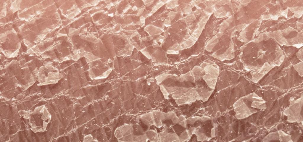
Varieties and classification of fish scale
There are several types of fish scale, which differ in their mode of inheritance, age of onset of skin lesions, clinical presentation, histopathological changes, and enzymatic and biochemical abnormalities. The most important subtypes include:
- common fish scale – occurs most often. Symptoms appear from about 3 to 12 months of age and localize primarily on the trunk and extremities. It can occur with atopic dermatitis,
- Hedgehog fish scales – involves lesions all over the skin, as well as skin folds and joint folds. It appears as early as birth or after birth,
- X-chromosome-coupled recessive fish scale,
- Arlequin fish scale – this is one of the most severe forms of the condition, which is characterized by various deformities in addition to excessive keratinization of the skin,
- classic fish scale – it features large, thick and dark scales, which can also occur on the articular flexures and neck,
- ichthyotic erythroderma,
- bullous fish scale,
- superficial fish blistering scales.
What is the diagnostic process?
The diagnostic process of fish scale involves several steps that help identify and confirm the disease and determine its form and severity. The first step is to take a detailed medical history. The doctor asks about the symptoms, their onset, intensity, and family history to determine if there have been similar cases among relatives. It is important to find out when the symptoms began and how they affect the patient's daily life. A thorough physical examination is then performed. The doctor evaluates the appearance of the skin, its texture, thickness and the presence of characteristic scales. He looks for typical signs of fish scales, such as excessive dryness of the skin, flaking and accumulation of dead skin. Additional laboratory tests, including analysis of skin samples, may be ordered to further confirm the diagnosis. A skin biopsy is also performed to evaluate the microscopic image of the skin and confirm the presence of fish scale features. To accurately diagnose fish scale and determine its form, genetic testing is necessary. To do this, the patient's DNA is analyzed to look for mutations in genes related to keratinization. It is also important to assess the patient's overall health and identify any related health problems.
Fish scales – treatment and patient care
As of today, no therapies have been developed to completely cure this rare disease. The most common is symptomatic treatment, which consists of skin care, moisturization and hydration using special creams and ointments with urea and salicylic acid. Pharmacological treatment is based on taking steroids. In some cases, surgical treatment is a good option. In the care of patients with fish scales, regular and meticulous care of the patient's skin is fundamental. Skin lesions should be monitored closely, so that dermatological abnormalities can be detected quickly if necessary. After the child is discharged from the hospital, a dermatologist, ophthalmologist, ENT specialist and physiotherapist should be consulted regularly.
Conclusion
Living with fish scale is challenging, but understanding it can guide better care. This genetic skin disorder disrupts keratinization, leading to scaly, thickened skin and discomfort. While there’s no cure, early diagnosis, consistent skin care, and personalized treatment can greatly improve quality of life. From tailored moisturizers to ongoing medical support, managing symptoms allows patients to regain control. Knowledge empowers action—take steps to address the condition and support better health outcomes.